-
Courses

Courses
Choosing a course is one of the most important decisions you'll ever make! View our courses and see what our students and lecturers have to say about the courses you are interested in at the links below.
-
University Life
University Life
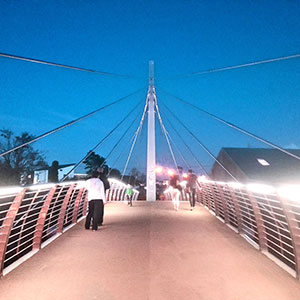
Each year more than 4,000 choose University of Galway as their University of choice. Find out what life at University of Galway is all about here.
-
About University of Galway
About University of Galway
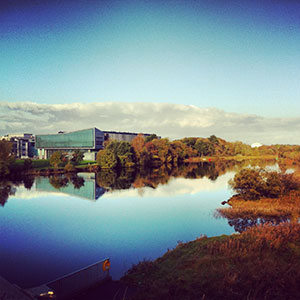
Since 1845, University of Galway has been sharing the highest quality teaching and research with Ireland and the world. Find out what makes our University so special – from our distinguished history to the latest news and campus developments.
-
Colleges & Schools
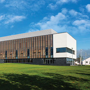
Colleges & Schools
University of Galway has earned international recognition as a research-led university with a commitment to top quality teaching across a range of key areas of expertise.
-
Research & Innovation

Research & Innovation
University of Galway’s vibrant research community take on some of the most pressing challenges of our times.
-
Business & Industry
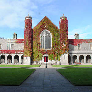
Guiding Breakthrough Research at University of Galway
We explore and facilitate commercial opportunities for the research community at University of Galway, as well as facilitating industry partnership.
-
Alumni & Friends

Alumni & Friends
There are 128,000 University of Galway alumni worldwide. Stay connected to your alumni community! Join our social networks and update your details online.
-
Community Engagement
Community Engagement
At University of Galway, we believe that the best learning takes place when you apply what you learn in a real world context. That's why many of our courses include work placements or community projects.
Instrumentation
Instrumentation
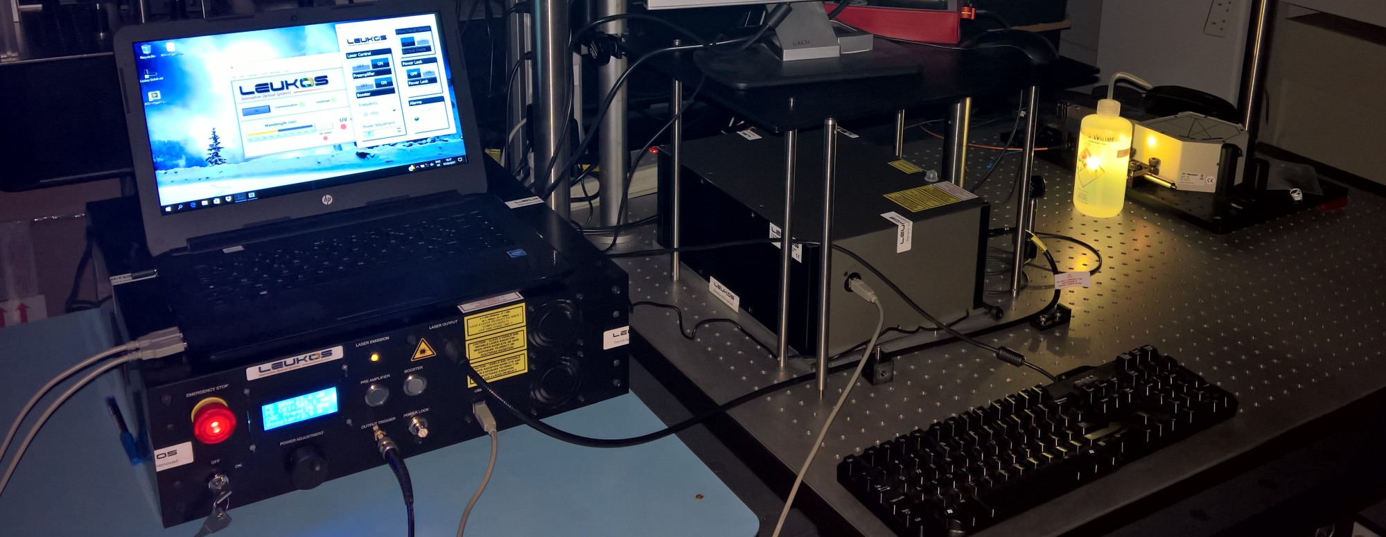
The NBL maintains and operates a very comprehensive suite of advanced spectroscopic instrumentation to further our research goals. This includes:
- An extensive array of benchtop spectrometers for UV-visible, fluorescence, and Raman spectroscopy.
- A cohort of HPLC, DLS, water activity, refractometers, viscometer, and other general analytical instrumentation.
- A suite of confocal fluorescence and Raman microscopes, laser sources, and advanced data collection and analysis hardware.
The facilities are available to research collaborators and for industrial consultancy subject to discussion and fee agreements where necessary.
General Analytical Instrumentation:
The group has a wide variety of general analytical instrumentation for elemental and molecular analysis. Two HPLC systems (Waters & Agilent 1260) are available for chromatographic analysis of small and large molecules. A water activity measurement system (AquaLab 4TE) and an osmometer (Osmomat 3000, Gonotech) are also available and typically used for cell culture media analysis. Two Dynamic Light Scattering systems are available for nanoparticle size analysis. We also have a refractometer and a cone-plate viscometer for the analysis of high concentration protein formulations.
Our elemental analysis facilities include an X-Ray Fluorescence spectrometer, a microwave plasma atomic emission spectrometer (MP-4200, from Agilent), and several ion selective electrodes.
Raman & FT-IR facilities:
The Raman page details the vibrational spectroscopy instrumentation available.
Currently we have four Raman spectrometers in operation and two FT-IR with ATR (Cary 630).
Fluorescence spectroscopy:
The NBL has nine steady state (2 x Horiba Aqualog, 4 x Cary Eclipse, 1 x LS50B, and 2 x Ocean optics) and four fluorescence lifetime based systems (3 x PicoQuant, and 1 x Becker & Hickl) for general spectroscopy.
Full details are available on the Fluorescence page.
Advanced microscopy:
The group currently has two confocal and one TIRF microsocopy systems in operation.
We also have a wide variety of optical components and additional spectrometers for adding to the microscopes as required. Full details are available on the Microscopy page.


